Receptor tyrosine kinase signaling pathway
Receptor tyrosine kinases (RTKs) dimerize upon ligand engagement, trans-autophosphorylate cytoplasmic kinase domains, and recruit SH2/PTB adaptors to couple Ras–Raf–MEK–ERK, PI3K–Akt, mTORC1, and PLCγ programs controlling proliferation, survival, migration, and metabolism. Cancers often select ligand-independent signaling via kinase-domain mutations, amplifications, or fusions, shaping bypass and acquired resistance.
1. Key targets
Core nodes
- EGFR / ERBB2 / ERBB3 / ERBB4
ErbB family; EGFR/HER2 amplification or mutations in epithelial cancers; dimerization biases outputs.
- MET / HGF
Stromal–epithelial crosstalk, invasion, bypass resistance; exon 14 skipping alters turnover.
- ALK / ROS1 / RET / TRK(NTRK)
Fusion-driven druggable kinases; partner genes and histology inform testing and therapy.
Vascular & stroma RTKs
- VEGFR1/2/3 (FLT1, KDR, FLT4)—see angiogenesis hub
- PDGFRA/B, KIT (CD117)—stromal programs & drug response
Negative regulation & trafficking
- Phosphatases (e.g., PTPRJ), E3 ligases (CBL), endocytic turnover
- PI3K–PTEN and Ras–MAPK feedback shape signal duration
Supplement: RTK-related targets (gene symbols)
Below the overview, HGNC-style symbols group ErbB, MET/FGF, vascular/stromal RTKs, insulin-receptor family, fusion-prone kinases, adaptors with Ras–MAPK/PI3K coupling, and negative regulation/endocytosis. The RTK universe is large—this is a research-oriented subset; validate fusion breakpoints, isoforms, and expression with databases/papers.
ErbB / HER & ligands
ErbB / HER receptor tyrosine kinases
EGFR, ERBB2, ERBB3, ERBB4
Major ErbB ligands (representative)
Paracrine/autocrine context sets heterodimer bias.
EGF, TGFA, AREG, EREG, HBEGF, NRG1, NRG2, NRG3, NRG4, BTC
MET & FGFR axes
MET / HGF axis
MET, HGF
FGFR & FGF (representative)
Developmental vs oncogenic hotspots differ; mind splice isoforms.
FGFR1, FGFR2, FGFR3, FGFR4, FGF1, FGF2, FGF3, FGF4, FGF5, FGF6, FGF7, FGF8, FGF9, FGF10, FGF18, FGF23
Vascular, stromal & myeloid RTKs
VEGFR family
See site angiogenesis hub for broader vascular programs.
FLT1, KDR, FLT4
PDGFR, KIT, CSF1R (stroma/myeloid)
PDGFRA, PDGFRB, KIT, CSF1R
TIE receptors & angiopoietins
TEK, TIE1, ANGPT1, ANGPT2
Insulin receptor family
Insulin receptor family
Metabolism–growth intersection; IGF2 can signal via INSR isoforms/hybrid receptors.
INSR, IGF1R, INSRR
Fusion-prone / diagnostic RTKs
ALK / ROS1 / RET
ALK, ROS1, RET
NTRK (Trk) family
NTRK1, NTRK2, NTRK3
FLT3 & other kinase targets (representative)
FLT3, DDR1, DDR2, MUSK
Adaptors & downstream branchpoints
SH2/PTB adaptors & scaffolds
GRB2, GRB7, SHC1, SHC2, SHC3, SHC4, GAB1, GAB2, IRS1, IRS2, FRS2, PTPN11
Ras–MAPK coupling (SOS/Ras switching)
See MAPK/ERK pathway page for cascade detail.
SOS1, SOS2, HRAS, KRAS, NRAS, RAF1, BRAF, MAP2K1, MAP2K2, MAPK1, MAPK3
PI3K & PLCγ branch nodes
Class I PI3K regulatory/catalytic subunits detailed on PI3K/Akt/mTOR page.
PIK3CA, PIK3CB, PIK3R1, PIK3R2, AKT1, AKT2, PLCG1, PLCG2
Negative regulation & endocytosis
E3 ligases & phosphotyrosine phosphatases
CBL, CBLB, PTPRJ, PTPRB, PTPRG, DUSP6, DUSP7
Endocytosis & trafficking (representative)
EPS15, EPN1, RAB5A, RAB7A
2. Suggested experimental readouts
Pair phospho-antibodies with totals and titrated stimulation; for fusions, complement IHC with breakpoint-aware PCR/NGS and expression controls.
- p-EGFR (sites per clone), total EGFR; surface levels and degradation kinetics
- p-MET (e.g., Y1234/1235), total MET; HGF co-culture stimulation
- p-ERK1/2, p-AKT (Ser473), p-S6 to separate MAPK vs PI3K branches
- ALK/ROS1/TRK fusions: breakpoint-aware reagents with diagnostic-grade controls
- Pair proliferation (Ki67) with apoptosis (cleaved caspase-3) where applicable
3. Ligand-driven activation vs genomic alterations
| Mode | Mechanism | Readouts / testing | Notes |
|---|---|---|---|
| Physiologic / autocrine | Ligand→dimerization→autophosphorylation→adaptors | Ligand trap, receptor blockade, pY readouts | Watch batch variability of serum growth factors. |
| Mutation / amplification | Conformational activation or overexpression lowers ligand threshold | NGS/ddPCR, FISH, IHC 3+ | Multiple resistance alleles can co-exist subclonally. |
| Fusion | Oligomerization domain–kinase constitutive activity | RNA-seq / FISH / IHC | Partner genes alter localization and turnover. |
4. Disease & model context
Development, repair & metabolism (non-oncology)
- FGFR signaling shapes limb, cranial, and urogenital development; gain-of-function alleles link to syndromic phenotypes—separate embryonic vs adult tissue models.
- PDGFR/KIT programs operate in wound healing, fibrosis, and mesenchymal activation—pair readouts with TGF-β and inflammatory cytokines.
- INSR/IGF1R couples nutrient sensing, growth, and insulin sensitivity—basal phosphotyrosine backgrounds are serum- and dose-sensitive.
Oncology: drugs, bypass, resistance
- Tyrosine kinase inhibitors (TKIs): ATP-competitive pocket occupancy; mutation spectra set sensitivity vs resistance (e.g., EGFR T790M, ALK gatekeeper).
- Bypass activation: MET, ERBB3, FGFR, AXL, etc., can compensate when primary RTKs are inhibited.
- Antibody therapeutics: Extracellular blockade, ADCC; combination design per guidelines and ethics.
RTK-related antibodies (curated)
5. Product lines & on-site search
Primary & phospho antibodies
EGFR, p-EGFR, ERBB2/HER2, MET, p-MET, ALK, ROS1, RET, TRK, KIT, FLT3, PDGFRA, VEGFR2—WB/IHC/IF/FC per clone/species.
Recombinant proteins / cytokines
EGF, HGF, PDGF-BB, VEGF for receptor stimulation—match species and endotoxin controls.
Functional reagents & quantification
Neutralizing antibodies, ECD-Fc traps, phospho/total ELISAs (availability-dependent); follow ethics/biosafety.
Quick search (site)
6. Inhibitors & tool compounds (summary)
For research use; follow lab SOP and datasheets. Clinical agents depend on institutional access.
EGFR TKIs; common in sensitizing-mutation models.
Third-generation EGFR TKI; T790M/C797S resistance studies.
Dual EGFR/ERBB2 TKI; breast-cancer readouts.
ALK/ROS1/MET (dose-dependent) inhibitor; fusion model control.
Pan-TRK/ALK/ROS1 (spectrum-dependent); NTRK fusion studies.
Multi-kinase TKIs including VEGFR/MET; mind off-targets.
BCR-ABL/KIT/PDGFRA; common positive control beyond classical RTKs.
Combo for MAPK-feedback experiments (project-dependent).
7. Pathway schematic
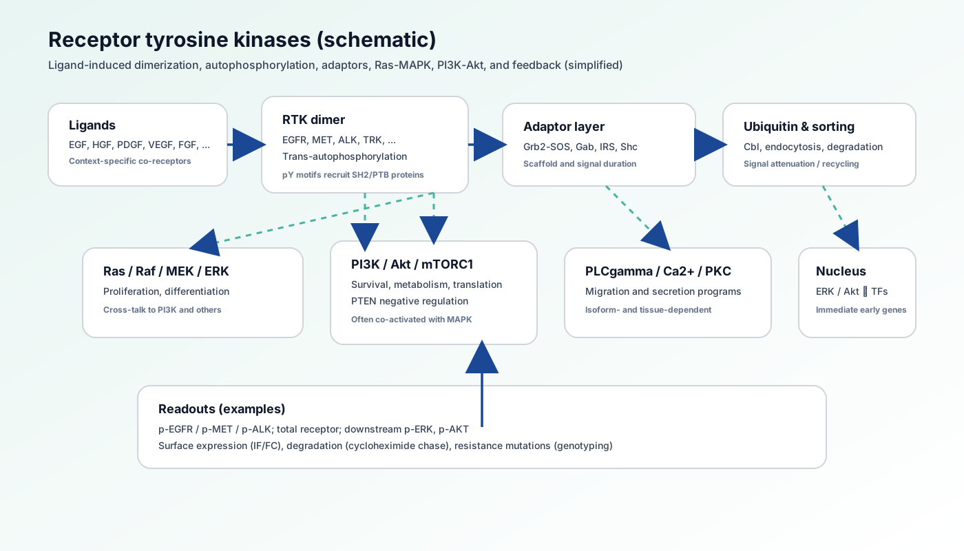
- Ligand-induced dimerization and trans-autophosphorylation
- SH2 adaptors (e.g., Grb2–SOS, Shc) couple Ras·GTP loading to MAPK cascades
- PI3K regulatory subunits read pY motifs for PIP3–Akt signaling
- PLCγ–Ca²⁺–PKC arms support migration/secretion
- Trafficking, ubiquitination, and phosphatases close loops (pair with Table 3)
8. Pathway biology overview
RTK networks coordinate intercellular communication in development, homeostasis, and repair; pathologically, genomic lesions plus microenvironmental ligands elevate baseline phosphotyrosine signaling, enabling plastic invasion and drug escape.
- Cells can co-depend on multiple RTKs—use multiplexed readouts
- TKIs vs antibodies act at distinct nodes—do not interchange conclusions
9. Related pathways on this site
10. External databases & modification resources
- KEGG: hsa04012 ErbB signaling pathway
- KEGG: hsa04014 Ras signaling pathway
- KEGG: hsa04010 MAPK signaling pathway (RTK→Ras coupling)
- Reactome: Signaling by EGFR
- Reactome: Signaling by MET
- Reactome: Signaling by Receptor Tyrosine Kinases (overview)
- Reactome: Signaling by FGFR
- WikiPathways (search: receptor tyrosine kinase)
- PhosphoSitePlus (RTK tyrosine phosphorylation sites)
11. References
RTK structure & signaling principles
- • Ullrich A, Schlessinger J. (1990). Cell. 61(2):203-12.
- • Lemmon MA, Schlessinger J. (2010). Cell. 141(7):1117-34.
Oncology targeting & resistance
- • Lynch TJ, et al. (2004). N Engl J Med. 350(21):2129-39.
- • Kwak EL, et al. (2010). N Engl J Med. 363(18):1693-703.