Angiogenesis signaling
Neovascularization couples hypoxia-inducible transcription, VEGF–VEGFR–driven endothelial proliferation/migration, Notch/DLL4 tip–stalk fate decisions, and Tie2–angiopoietin control of maturation and pericyte coverage. Pathological vessels in cancer, chronic inflammation, or ischemia often show high permeability, structural immaturity, and therapy resistance—interpret with multiplex readouts.
1. Key targets
Core nodes
- VEGFA / VEGFC & KDR / FLT1
Ligand–receptor tyrosine kinase axis; sprouting, survival, permeability (context-dependent).
- TEK (Tie2) / ANGPT1 / ANGPT2
Vessel maturation/stability; ANGPT2 often destabilizes in tumor settings.
- NOTCH1 / DLL4 / JAG1
DLL4-high tip cells and lateral inhibition; branching patterning.
Hypoxia & transcription
- HIF1A / EPAS1 (HIF2A); PHD–VHL regulation
- Downstream clusters (VEGFA, GLUT1, LDHA—model-dependent)
Endothelial phenotype & matrix
- CDH5 (VE-cadherin), CD31, Endoglin (ENG)
- MMP2/9, integrins, basement-membrane collagen remodeling
- PDGFRB, NG2 (CSPG4)—pericyte coverage
2. Suggested experimental readouts
Pair structural readouts (pericytes, basement membrane) with functional assays (perfusion, permeability); for phospho targets, include totals and dose/time controls.
- CD31 / CD34 / IB4 with sprout metrics (organoid/aortic ring models)
- p-ERK, p-AKT; total KDR/FLT1 (phospho sites per clone)
- DLL4, NOTCH1 enrichment at tips; Hey1/HEY2 transcriptional readouts
- α-SMA / PDGFRB / NG2 with CD31—pericyte coverage index
- Hypoxia probes or HIF1A nuclear localization (fixation-sensitive)
- Tubulogenesis assays (batch-to-batch matrix/serum variability)
3. VEGF axis vs Tie2–angiopoietin axis
| Axis | Key ligand–receptor | Typical roles | Practical notes |
|---|---|---|---|
| VEGF | VEGFA/C → KDR / FLT1 | Sprouting, migration, survival, permeability | TKIs vs neutralizing antibodies differ mechanistically. |
| Angpt–Tie2 | ANGPT1/2 → TEK (Tie2) | Maturation/stability; tumor contexts remodel mural attachment | Pair with VEGF for normalization vs resistance narratives. |
4. Tumor vessels: phenotypes, normalization & resistance
- Immature structure: Discontinuous basement membrane, low pericyte coverage—often high leakiness and interstitial pressure.
- Normalization window: Transient perfusion/delivery improvements; timing is tumor- and regimen-specific.
- Compensatory pathways: FGF, Angpt, Notch shifts, or myeloid/stromal reprogramming can drive revascularization.
Angiogenesis-related antibodies (curated)
5. Product lines & on-site search
Primary & phospho antibodies
VEGFA, KDR, FLT1, Tie2, DLL4, Notch1, CD31, CDH5, Endoglin, HIF1A, MMP2/9, PDGFRB—WB/IHC/IF/FC as applicable.
Recombinant proteins / cytokines
VEGF165, PlGF, Angpt1/2 for co-culture/stimulation—match species and titrate controls.
Functional reagents & quantification
Neutralizing anti-VEGF, soluble traps, species-matched VEGFA ELISAs—follow datasheets and ethics.
6. Inhibitors & tool compounds (summary)
For research use; follow lab SOP and datasheets. Clinical biologics (e.g., bevacizumab-class) depend on institutional access.
Multi-kinase inhibitor; VEGFR/PDGFR/c-KIT (model-dependent).
RAF/VEGFR multi-target; common control arm.
VEGF-axis-preferring TKI; common in vascular phenotyping.
Broader VEGFR/FGFR profile; watch off-target toxicity.
VEGFR1–3 / FGFR / PDGFR, etc.; combine studies need monotherapy controls.
MET/VEGFR2, etc.; invasion–angiogenesis crosstalk studies.
VEGFR2-selective small molecule (regional approval varies).
γ-secretase / Notch tools; alters tip–stalk allocation.
7. Pathway schematic
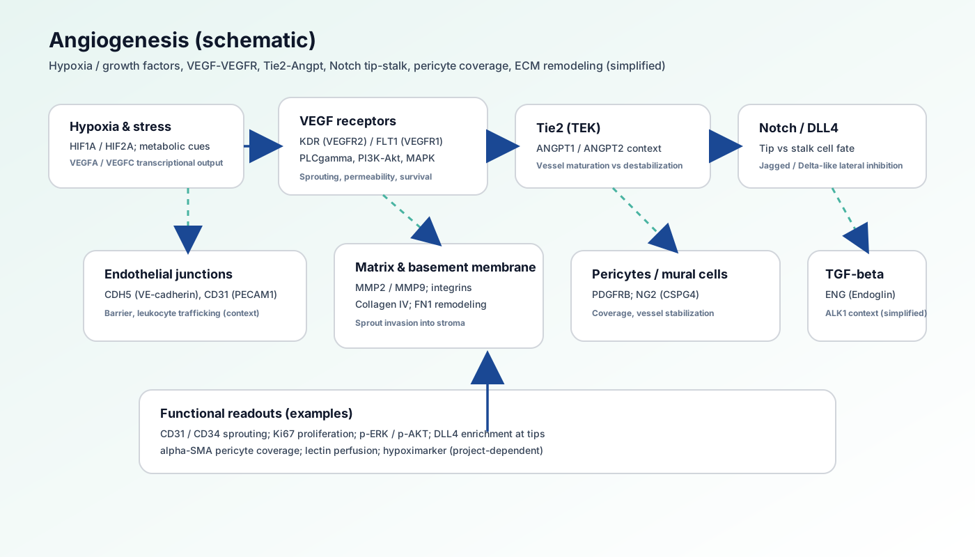
- Hypoxia–HIF drives VEGF transcriptional output
- VEGFR→MAPK/PI3K and migratory cytoskeleton programs
- Tie2–Angpt regulates stability and mural attachment
- Notch/DLL4 with matrix remodeling shapes branching
8. Pathway biology overview
Physiologic angiogenesis is tightly spatiotemporally controlled during development and repair; pathology couples pro-angiogenic cues, matrix mechanics, and immune cells to reprogram endothelial metabolism and heterogeneity.
- Lymphangiogenesis (VEGFC/D–FLT4) may parallel blood vessels—use distinct markers
- Organ specificity: BBB vs liver sinusoids differ substantially
9. Related pathways on this site
10. External databases & modification resources
- KEGG: hsa04370 VEGF signaling pathway
- Reactome: Signaling by VEGF
- WikiPathways (search: angiogenesis)
- PhosphoSitePlus (receptor tyrosine phosphorylation sites)
11. References
VEGF & angiogenesis mechanisms
- • Ferrara N, Kerbel RS. (2005). Nature. 438(7070):967-74.
- • Carmeliet P, Jain RK. (2011). Nature. 473(7347):298-307.
Tip cells & Notch
- • Hellström M, et al. (2007). Nature. 445(7129):776-80.
- • Phng LK, Gerhardt H. (2009). J Cell Sci. 122(Pt 17):3151-9.