Cellular autophagy signaling
Cellular autophagy most often denotes macroautophagy: cytoplasm is sequestered in double-membrane autophagosomes that fuse with lysosomes for catabolic recycling. The ULK complex and class III PI3K (VPS34–Beclin) nucleate/elongate the phagophore; ATG12–ATG5–ATG16L1 with ATG7/ATG3 lipidates LC3 (MAP1LC3) to LC3-II. Starvation, mTORC1 inhibition, and AMPK activation are common inducers; selective autophagy couples ubiquitin to receptors (mitophagy, aggrephagy, etc.). Programs can be homeostatic, tumor-suppressive, or pro-survival depending on context.
1. Key targets
Initiation & nucleation
- ULK1 / ULK2
ULK1/2 with FIP200 (RB1CC1), ATG13, ATG101; regulated by mTORC1 and AMPK phosphorylation.
- PIK3C3 / VPS34
Class III PI3K generates PI(3)P; Beclin-1 partners tune complex outputs.
- WIPI1 / WIPI2
PI(3)P effectors linking ATG machinery to membrane expansion.
Elongation & closure
- ATG12–ATG5 偶联;ATG16L1 定位于新生膜边缘
- ATG7/ATG3 conjugate LC3-PE (LC3-II)
Fusion & cargo
- LAMP1 / LAMP2、Rab7、SNARE 介导自噬体–溶酶体融合
- p62/SQSTM1, NBR1, OPTN—selective receptors
Supplement: Autophagy targets (gene symbols)
HGNC symbols for ULK, Beclin, ATG conjugation, lysosome, and selective autophagy modules.
Conjugation & LC3
2. Suggested experimental readouts
Interpret LC3-II with lysosomal inhibitors (BafA1/CQ) for flux; p62 alone is not sufficient.
- LC3A/B and GABARAP isoforms; LC3-II/LC3-I with inhibitor chase
- p62, NBR1; pair with ubiquitinated cargo
- Beclin-1, ATG5/7, ULK1 total/phospho (clone-dependent)
- LAMP1/2 with LC3 colocalization (IF)
- Mitophagy: PINK1, Parkin, mitochondrial markers
3. Autophagy modalities (brief)
| Type | Membrane / compartment | Example nodes |
|---|---|---|
| Macroautophagy | Autophagosome (double membrane) | ULK, VPS34–Beclin, ATG12–ATG5–ATG16L1, LC3-II |
| CMA | Hsc70; LAMP2A translocation | HSPA8, LAMP2A, KFERQ-like clients |
| Microautophagy | Lysosomal invagination | Highly cargo- and cell-type–dependent |
4. Physiology & disease context
- Metabolism & infection: amino-acid/lipid recycling; pathogens can hijack or evade autophagy membranes.
- Neurodegeneration: aggregate clearance and axonal autophagy flux are frequent themes.
- Cancer: stress tolerance vs early tumor suppression—crosstalk with mTOR/MAPK.
Cellular autophagy–related antibodies (curated)
5. Product lines & on-site search
Primary & phospho antibodies
LC3, p62, Beclin-1, ATG family, ULK1, LAMP1/2, PINK1, Parkin, OPTN, TBK1—WB/IF/EM per datasheet.
Functional & tracer reagents
LysoTracker-class dyes, mCherry-GFP-LC3 reporters (availability-dependent); compounds per ethics.
6. Inhibitors & tool compounds (summary)
Clinical agents are prescription drugs; research use requires datasheets and compliance.
V-ATPase blockade; LC3-II flux pairing.
Lysosomotropic weak bases—context differs from clinic.
PI3K-family inhibition—class III vs I off-targets.
mTOR inhibition—common autophagy induction lever.
VPS34- or USP-axis probes for initiation.
Depolarization for mitophagy models—toxicity controls.
7. Pathway schematic
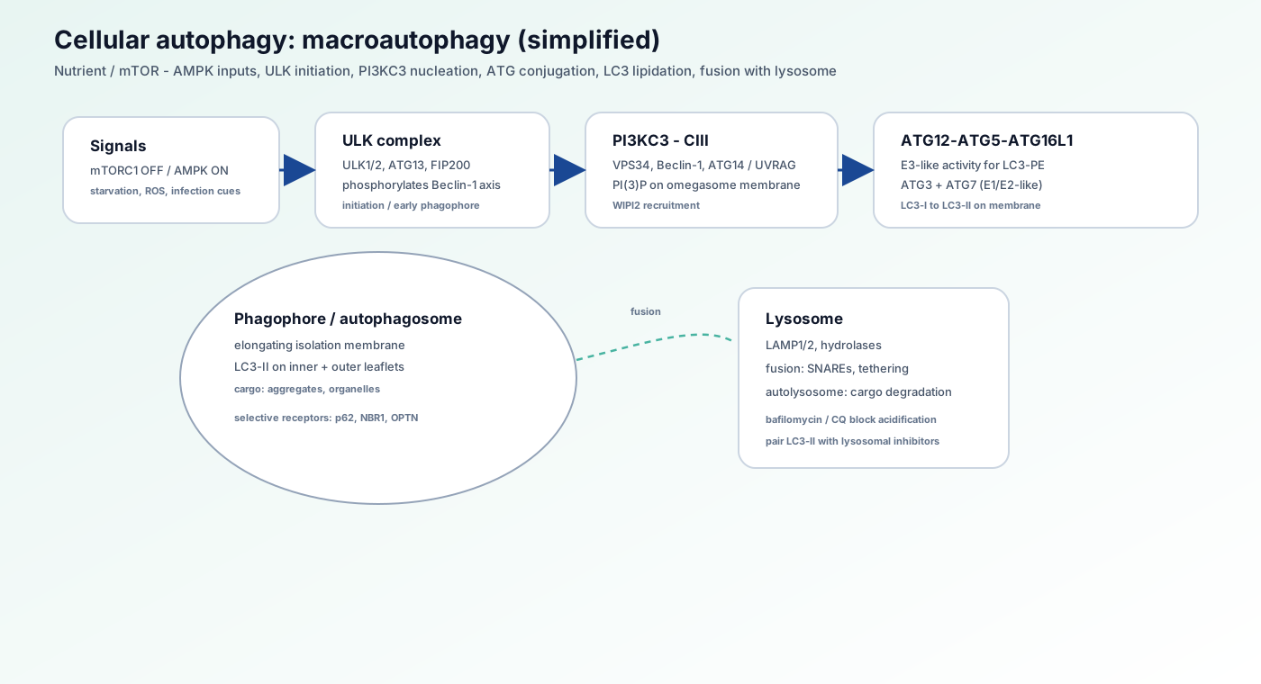
- Nutrient cues tune ULK via mTOR/AMPK
- PI(3)P/WIPI recruit ATG machinery
- LC3-II marks expanding membranes
- Lysosomal fusion completes degradation
8. Pathway biology overview
Cellular autophagy is a major degradative/recycling route that cooperates with the proteasome for proteostasis and organelle quality. Activity integrates nutrient, energy, redox, and infection cues; flux—not a single marker level—is often the key functional readout.
- Immunity: cross-presentation, pathogen interplay
- Development: spatiotemporal autophagy in remodeling
9. Related pathways on this site
10. External databases & guidelines
11. References
Core reviews
- • Mizushima N, Komatsu M. (2011). Autophagy: renovation of cells and tissues. Cell. 147(4):728-41.
- • Levine B, Kroemer G. (2008). Autophagy in the pathogenesis of disease. Cell. 132(1):27-42.
Assay guidance
- • Klionsky DJ, et al. (2021). Guidelines for the use and interpretation of assays for monitoring autophagy (4th edition). Autophagy. 17(1):1-382.